MechoApedia by KREATiS

◄ back

MechoApedia

close

Description

MechoApedia is a tool dedicated to learning the scientific basis of the mechanisms of toxic action, termed "MechoA" by KREATiS, classified within this scheme. MechoAs are specific molecular initiating events, the first step in Adverse Outcome Pathways, responsible for toxicity to biological organisms of all kinds and therefore these classifications are relevant to both ecotoxicologists and human health specialists (Bauer et al., 2018a, 2018b). This literature review is taken from Bauer’s thesis on MechoAs (Bauer, 2017). Click on MechoAs and their subclasses to get specific information on the interactions between the test substance and the biological matrices.


References:

Adams, W.J., Biddinger, G.R., Robillard, K.A., and Gorsuch, J.W. (1995). A summary of the acute toxicity of 14 phthalate esters to representative aquatic organisms. Environmental Toxicology and Chemistry 14, 1569–1574.

Airaksinen, M.M., Rosenberg, P.H., and Tammisto, T. (1970). A Possible Mechanism of Toxicity of Trifluoroethanol and Other Halothane Metabolites. Acta Pharmacologica et Toxicologica 28, 299–304.

ATSDR (1997a). Toxicological Profile: Chloroform.

ATSDR (1997b). Toxicological Profile: Hydrazines.

ATSDR (2014). Medical Management Guidelines (MMGs): Methylene Chloride.

Barron, A.B., Søvik, E., and Cornish, J.L. (2010). The Roles of Dopamine and Related Compounds in Reward-Seeking Behavior Across Animal Phyla. Front. Behav. Neurosci. 4.

Bauer, F. (2017). Une meilleure caractérisation des mécanismes d’action toxique à partir de la structure moléculaire. Université de Haute-Alsace.

Bauer, F.J., Thomas, P.C., Fouchard, S.Y., and Neunlist, S.J.M. (2018a). A new classification algorithm based on mechanisms of action. Computational Toxicology 5, 8–15.

Bauer, F.J., Thomas, P.C., Fouchard, S.Y., and Neunlist, S.J.M. (2018b). High-accuracy prediction of mechanisms of action using structural alerts. Computational Toxicology 7, 36–45.

Berg, J.M., Tymoczko, J.L., and Stryer, L. (2002). A Proton Gradient Powers the Synthesis of ATP. In Biochemistry, (New-York: W.H. Freeman), p.

Blanco-Ayala, T., Andérica-Romero, A.C., and Pedraza-Chaverri, J. (2014). New insights into antioxidant strategies against paraquat toxicity. Free Radical Research 48, 623–640.

Bolton, J.L., Trush, M.A., Penning, T.M., Dryhurst, G., and Monks, T.J. (2000). Role of Quinones in Toxicology. Chemical Research in Toxicology 13, 135–160.

Brown, M.A., and Vito, S.C.D. (1993). Predicting azo dye toxicity. Critical Reviews in Environmental Science and Technology 23, 249–324.

Brüning, T., and Bolt, H.M. (2000). Renal toxicity and carcinogenicity of trichloroethylene: key results, mechanisms, and controversies. Crit. Rev. Toxicol. 30, 253–285.

Calipari, E.S., and Ferris, M.J. (2013). Amphetamine Mechanisms and Actions at the Dopamine Terminal Revisited. J Neurosci 33, 8923–8925.

Casey, J.R., Grinstein, S., and Orlowski, J. (2010). Sensors and regulators of intracellular pH. Nat Rev Mol Cell Biol 11, 50–61.

Chahl, L.A. (1996). Experimental and Clinical Pharmacology: Opioids - mechanisms of action. Australian Prescriber 19, 63–65.

Coats, J.R. (1990). Mechanisms of toxic action and structure-activity relationships for organochlorine and synthetic pyrethroid insecticides. Environ Health Perspect 87, 255–262.

Committee on Acute Exposure Guideline Levels, Committee on Toxicology, Board on Environmental Studies and Toxicology, Division on Earth and Life Studies, and National Research Council (2012). Piperidine. In Acute Exposure Guideline Levels for Selected Airbone Chemicals, (National Academies Press (US)), p.

Dearden, J.C. (2002). Prediction of environmental toxicity and fate using quantitative structure-activity relationships (QSARs). Journal of the Brazilian Chemical Society 13, 754–762.

Di Francesco, A.M., Ward, T.H., and Butler, J. (2004). Diaziridinylbenzoquinones. In Quinones and Quinone Enzymes, (Academic Press), pp. 181–182.

Droual, B. (2011). Chapter 8 - Synaptic Transmission and Neural Integration.

Dunn, M.F., Ramírez-Trujillo, J.A., and Hernández-Lucas, I. (2009). Major roles of isocitrate lyase and malate synthase in bacterial and fungal pathogenesis. Microbiology 155, 3166–3175.

Elersek, T., and Filipic, M. (2011). Organophosphorus Pesticides - Mechanisms of Their Toxicity. In Pesticides - The Impacts of Pesticides Exposure, M. Stoytcheva, ed. (InTech), p.

Ellison, C.M., Madden, J.C., Cronin, M.T.D., and Enoch, S.J. (2015a). Investigation of the Verhaar scheme for predicting acute aquatic toxicity: Improving predictions obtained from Toxtree ver. 2.6. Chemosphere 139, 146–154.

Ellison, C.M., Madden, J.C., Cronin, M.T.D., and Enoch, S.J. (2015b). Investigation of the Verhaar scheme for predicting acute aquatic toxicity: Improving predictions obtained from Toxtree ver. 2.6. Chemosphere 139, 146–154.

Endo, S., Brown, T.N., and Goss, K.-U. (2013). General Model for Estimating Partition Coefficients to Organisms and Their Tissues Using the Biological Compositions and Polyparameter Linear Free Energy Relationships. Environ. Sci. Technol. 47, 6630–6639.

Enoch, S.J., Hewitt, M., Cronin, M.T.D., Azam, S., and Madden, J.C. (2008). Classification of chemicals according to mechanism of aquatic toxicity: An evaluation of the implementation of the Verhaar scheme in Toxtree. Chemosphere 73, 243–248.

Escher, B.I., and Hermens, J.L.M. (2002). Modes of Action in Ecotoxicology:  Their Role in Body Burdens, Species Sensitivity, QSARs, and Mixture Effects. Environ. Sci. Technol. 36, 4201–4217.

Escher, B.I., Hunziker, R., Schwarzenbach, R.P., and Westall, J.C. (1999). Kinetic Model To Describe the Intrinsic Uncoupling Activity of Substituted Phenols in Energy Transducing Membranes. Environ. Sci. Technol. 33, 560–570.

Food and Drug Administration (2004). Docket No. 2004N-0205 Furan in Food, Thermal Treatment; Request for Data and Information.

Franks, N.P., and Lieb, W.R. (1978). Where do general anaesthetics act? Nature 274, 339–342.

Franks, N.P., and Lieb, W.R. (1998). Which molecular targets are most relevant to general anaesthesia? Toxicology Letters 100–101, 1–8.

Fukuto, T.R. (1990). Mechanism of action of organophosphorus and carbamate insecticides. Environ Health Perspect 87, 245–254.

Grogan, J., DeVito, S.C., Pearlman, R.S., and Korzekwa, K.R. (1992). Modeling cyanide release from nitriles: prediction of cytochrome P 450-mediated acute nitrile toxicity. Chem. Res. Toxicol. 5, 548–552.

IAC Publishing Labs How does caffeine affect plant growth?

Ioannides, C. (1996). The CYP2E Subfamily - Evolution. In Cytochromes P450: Metabolic and Toxicological Aspects, (CRC Press), p. 213.

IPCS Inchem (1998). Diphenylamine (addendum). In JMPR Evaluations, (Rome), p.

IPCS Inchem (2002). Monograph on Atropine - Antidotes for poisoning by organophosphorus pesticides.

Jacobs, A. (1997). Hard and soft nucleophiles. In Understanding Organic Reaction Mechanisms, (Cambridge University Press), pp. 45–46.

Jaworska, J.S., Hunter, R.S., and Schultz, T.W. (1995). Quantitative structure-toxicity relationships and volume fraction analyses for selected esters. Arch. Environ. Contam. Toxicol. 29, 86–93.

Kazius, J., McGuire, R., and Bursi, R. (2005). Derivation and Validation of Toxicophores for Mutagenicity Prediction. J. Med. Chem. 48, 312–320.

King, M.W. (2015a). Ethanol (Alcohol) Metabolism: Acute and Chronic Toxicities.

King, M.W. (2015b). Biochemistry of Neurotransmitters and Nerve Transmission.

Koleva, Y., and Barzilov, I. (2010). Comparative study of mechanism of action of allyl alcohols for different endpoints. 49, 55–59.

Kulma, A., and Szopa, J. (2007). Catecholamines are active compounds in plants. Plant Science 172, 433–440.

Li, X. (2009). Glutathione and Glutathione-S-Transferase in Detoxification Mechanisms. In General, Applied and Systems Toxicology, (John Wiley & Sons, Ltd), p.

Liska, D.J. (1998). The detoxification enzyme systems. Altern Med Rev 3, 187–198.

Lugli, A.K., Yost, C.S., and Kindler, C.H. (2009). Anaesthetic mechanisms: update on the challenge of unravelling the mystery of anaesthesia. Eur J Anaesthesiol 26, 807–820.

McCarthy, M.C., and Enquist, B. (2005). Organismal size, metabolism and the evolution of complexity in metazoans. Evolutionary Ecology Research 7, 681–696.

Moridani, M.Y., Cheon, S.S., Khan, S., and O’Brien, P.J. (2003). Metabolic activation of 3-hydroxyanisole by isolated rat hepatocytes. Chemico-Biological Interactions 142, 317–333.

Munday, R. (1989). Toxicity of thiols and disulphides: Involvement of free-radical species. Free Radical Biology and Medicine 7, 659–673.

Nair, B. (2001). Final report on the safety assessment of Benzyl Alcohol, Benzoic Acid, and Sodium Benzoate. Int. J. Toxicol. 20 Suppl 3, 23–50.

National Center for Biotechnology Information (2017). CONIINE | C8H17N.

Nehlig, A., Daval, J.-L., and Debry, G. (1992). Caffeine and the central nervous system: mechanisms of action, biochemical, metabolic and psychostimulant effects. Brain Research Reviews 17, 139–170.

O’Donoghue, J.L. (2001). Ketones of Four or Five Carbons. In Patty’s Toxicology, (John Wiley & Sons, Inc.), p.

Olsen, R.W., and DeLorey, T.M. (1999). GABA Receptor Physiology and Pharmacology.

OpenStax (2014). Carbohydrate Metabolism. In Anatomy & Physiology, p.

Petering, D.H., and Fowler, B.A. (1986). Roles of metallothionein and related proteins in metal metabolism and toxicity: problems and perspectives. Environ Health Perspect 65, 217–224.

Recknagel, R.O., Glende Jr., E.A., Dolak, J.A., and Waller, R.L. (1989). Mechanisms of carbon tetrachloride toxicity. Pharmacology & Therapeutics 43, 139–154.

van Rensen, J.J.S. (1982). Molecular mechanisms of herbicide action near photosystem II. Physiologia Plantarum 54, 515–521.

Roberts, D.W., Roberts, J.F., Hodges, G., Gutsell, S., Ward, R.S., and Llewellyn, C. (2013). Aquatic toxicity of cationic surfactants to Daphnia magna. SAR and QSAR in Environmental Research 24, 417–427.

Russom, C.L., Bradbury, S.P., Broderius, S.J., Hammermeister, D.E., and Drummond, R.A. (1997). Predicting modes of toxic action from chemical structure: Acute toxicity in the fathead minnow (Pimephales promelas). Environmental Toxicology and Chemistry 16, 948–967.

Schultz, T.W. (1987). The use of the ionization constant (pKa) in selecting models of toxicity in phenols. Ecotoxicol. Environ. Saf. 14, 178–183.

Shaw, W. (2002). Chapter 3 Organic Acid Testing, Byproducts of Yeast and their Relationship to Autism. In Biological Treatments for Autism and PDD. Causes and Biomedical Therapies for Autism & PDD., p.

Sherer, T.B., Richardson, J.R., Testa, C.M., Seo, B.B., Panov, A.V., Yagi, T., Matsuno-Yagi, A., Miller, G.W., and Greenamyre, J.T. (2007). Mechanism of toxicity of pesticides acting at complex I: relevance to environmental etiologies of Parkinson’s disease. Journal of Neurochemistry 100, 1469–1479.

Sikkema, J., de Bont, J.A., and Poolman, B. (1995). Mechanisms of membrane toxicity of hydrocarbons. Microbiol Rev 59, 201–222.

Smith, G.F. (2011). Designing Drugs to Avoid Toxicity. In Progress in Medicinal Chemistry, G. Lawton, and D.R. Witty, eds. (Elsevier), pp. 1–47.

de Sousa, J.P., Torres, R. de A., de Azerêdo, G.A., Figueiredo, R.C.B.Q., Vasconcelos, M.A. da S., and de Souza, E.L. (2012). Carvacrol and 1,8-cineole alone or in combination at sublethal concentrations induce changes in the cell morphology and membrane permeability of Pseudomonas fluorescens in a vegetable-based broth. International Journal of Food Microbiology 158, 9–13.

Vaes, W.H., Ramos, E.U., Verhaar, H.J., and Hermens, J.L. (1998). Acute toxicity of nonpolar versus polar narcosis: Is there a difference? Environmental Toxicology and Chemistry 17, 1380–1384.

Vallabh Minikel, E. (2016). A mechanism of action hypothesis.

Veith, G.D., and Broderius, S.J. (1987). Structure-Toxicity Relationships for Industrial Chemicals Causing Type (II) Narcosis Syndrome. In QSAR in Environmental Toxicology - II, K.L.E. Kaiser, ed. (Springer Netherlands), pp. 385–391.

Verhaar, H.J.M., van Leeuwen, C.J., and Hermens, J.L.M. (1992). Classifying environmental pollutants. Chemosphere 25, 471–491.

Vetter, J. (2004). Poison hemlock (Conium maculatum L.). Food and Chemical Toxicology 42, 1373–1382.

Way, J.L., Leung, P., Cannon, E., Morgan, R., Tamulinas, C., Leong-Way, J., Baxter, L., Nagi, A., and Chui, C. (2007). The Mechanism of Cyanide Intoxication and its Antagonism - Ciba Foundation Symposium 140 - Cyanide Compounds in Biology - Way - Wiley Online Library. (D. Evered and S. Hamett), pp. 232–248.

Wessels, S., and Ingmer, H. (2013). Modes of action of three disinfectant active substances: A review. Regulatory Toxicology and Pharmacology 67, 456–467.

Wiley-VCH Verlag (2002a). Organic peroxides [MAK Value Documentation, 1992]. In The MAK-Collection for Occupational Health and Safety, (Wiley-VCH Verlag GmbH & Co. KGaA), p.

Wiley-VCH Verlag (2002b). Triethylene glycol [MAK Value Documentation, 2007]. In The MAK-Collection for Occupational Health and Safety, (Wiley-VCH Verlag GmbH & Co. KGaA), p.

close

1.1 Non-Polar Narcosis

Non-polar narcosis (MechoA 1.1), or "baseline toxicity", is assumed to be the least toxic of the known mechanisms of action for less hydrophobic substances (Verhaar et al., 1992). Generally, at comparable hydrophobicities or solubilities, non-polar narcotics are less toxic than substances with any other MechoA, as observed by (Dearden, 2002).

Non-polar narcotics englobe a wide range of chemical families such as hydrocarbons, ketones, ethers, aliphatic alcohols and their halogenated derivatives etc. and is thought to account for more than 50% of the organic chemical universe (Ellison et al., 2015a).

close

1.2 Polar Narcosis

In practice, the distinction between non-polar and polar narcosis may be ambiguous. For examples, some pyridines are considered to act with MechoA 1.1 while other pyridines are associated to MechoA 1.2. Some authors consider the separation between those two MechoA to be linked to polarizability of the molecules, or even their abilities to participate in strong hydrogen bonding (Roberts et al., 2013). Others have suggested the difference between MechoA 1.1 and 1.2 was a consequence of the use of the log KOW to quantify the equilibrium of the molecules between aqueous medium and cell membranes. With log KOW, octanol is assumed to represent membrane lipids. Some experiments have been performed by replacing octanol with DMPC thus normalising the toxic impact between so-called polar and non-polar compounds (Vaes et al., 1998). Finally, other authors explained the difference of toxicity of these two groups by a difference of partitioning between different compartments, which is not taken into account with log KOW (Endo et al., 2013; Escher and Hermens, 2002).

The polar narcosis (MechoA 1.2) is very similar to non-polar narcosis despite of the significant difference of toxicity observed for compounds with equivalent hydrophobicity (Escher and Hermens, 2002; Verhaar et al., 1992). Indeed, effects due to carvacrol (MechoA 1.2) are comparable to 1,8-cineole (MechoA 1.1) but they are observable at smaller concentrations (de Sousa et al., 2012).

This mechanism concerns mainly simple phenols and anilines, but also amines and nitrogen heterocycles (Ellison et al., 2015b).

close

1.3 Cationic Narcosis

Cationic narcosis (MechoA 1.3) is based on the same principle as polar narcosis however their potency is higher because the bond energies involved are stronger and more favourable for interactions between cationic compounds and phospholipids of the cell membranes. The positively charged moiety of cationic molecules stays at the surface of the membrane and can disrupt the charge distribution there (Roberts et al., 2013).

This mechanism especially concerns quaternary ammonium compounds.

Due to their relative high toxicity to unicellular organisms compared to other narcotics, quaternary ammonium compounds are often used as antimicrobials with a broad spectrum activity (Wessels and Ingmer, 2013).

close

1 Narcosis

Narcosis is a non-specific, non-reactive and reversible process that any organic molecule can exert on cellular organisms (Verhaar et al., 1992). The principle of this MechoA is based on a simple accumulation of substance in cell membranes thus disturbing their functions up to the loss of physical integrity. There is a recognised strong relationship between toxicity and octanol-water partition coefficient (KOW) which has led to the hypothesis that narcosis is related to destabilisation of lipid membranes. However, the effect may be due to the impairment of cell proteins (e.g. ion channel or G proteins) because their function depends on their environment and their spatial conformation (Franks and Lieb, 1998; Sikkema et al., 1995). Since cell membranes are hydrophobic components, the main property driving the intensity of the adverse outcome due to these toxic mechanisms of action is the hydrophobicity of molecules. This property gives an indication of the relative fraction of the test substance in the aqueous compartment (i.e. extracellular medium or cytosol) and the fraction accumulated within hydrophobic compartments like membranes (target lipid) but also lipidic tissue (storage lipid). The potential of the narcotic effect varies according to the different affinities of non-polar and polar compounds for the membranes.

The term "narcosis" here is the same as that used to describe rapid and reversible anaesthesia. Anaesthetic potential of molecules has been demonstrated to be correlated to their hydrophobicity according to the Meyer-Overton relationship (Lugli et al., 2009). That would indicate the mechanism of action leading to anaesthesia is the accumulation of molecules within the neural cell membranes. However, several studies on the anaesthesia mechanisms have suggested the efficiency of narcotics is rather due to specific protein targets (like ion channels) which have non-specific binding site (Franks and Lieb, 1978; Lugli et al., 2009). After exposition to carvacrol or 1,8-cineole, some cavities are visible in cell membranes causing the cytosol to leave the cell (cf. in opposite Figure). 1,8-cineole is a simple aliphatic ether with a MechoA 1.1 while carvacrol exerts MechoA 1.2, as a simple alkylphenol. These observations illustrate the narcotic impact of molecules.

Figure: Bacteria P. fluorescens observed with Transmission Electron Microscopy (left column) and with Scanning Electron Microscopy (right column); (a-b) control, (c-d) after exposure to carvacrol, (e-f) after exposure to 1,8-cineole (de Sousa et al., 2012).

close

2.1 Narcotic products

Veith and Broderius (1987) identified that simple esters lead to symptoms similar to those of narcosis. Moreover, these compounds are not very reactive in neutral aqueous medium. Since they observed an excess of toxicity compared to narcotic compounds, these authors then described the toxicity of esters as another form of narcosis they termed "esters narcosis". Russom et al. (1997) also used this category in their classification.

Nevertheless, in 1995, Jaworska et al. reported that, while esters behave as non-polar narcotic compounds to protozoa (Tetrahymena pyriformis), they were actually more toxic than non-polar narcotics to fish (Pimephales promelas). The authors explained this observation by making the assumption that protozoa do not, or barely, metabolise esters while fish esterase enzymes can hydrolyse esters very efficiently (Figure below).

Figure: enzymatic hydrolysis of esters. R1=H or alkyl, R2=alkyl.

This hydrolysis changes the equilibrium concentration between the external aqueous medium and the internal fish concentration since the ester internal concentration is decreased by the hydrolysis reaction. Thus, due to thermodynamic equilibrium, more ester will be transferred from external medium to fish tissues. Consequently, the fish might potentially be exposed to a higher amount of ester than if it wasn't metabolised, although this equilibrium may be determined by the hydrophobicity of the ester itself. Uptake of more hydrophobic substances by aquatic organisms may occur less rapidly than their metabolization once inside the body giving an impression of reduced substance toxicity. Moreover, the hydrolysis mechanism favours ester depuration from aquatic organisms since the hydrolysis products are less hydrophobic than the ester parent. The metabolites are also less toxic than the ester parent substance. For less hydrophobic esters, accumulation of the alcohol generated after hydrolysis combined with rapid uptake of the parent compound may exert a significant narcotic effect. Besides, the hydrolysis reaction produces acidity which may contribute to the disturbance of normal cell functioning (see MechoA 5.2).

close

2.2 Non-narcotic products

The hydrolysis product of esters is often an alcohol but not always. For a phenyl ester, for example, the degradation product will be a phenol with a more toxic MechoA than MechoA 1.1 resulting in various possible outcomes: polar narcosis (see MechoA 1.2); uncoupling of oxidative phosphorylation (see MechoA 5.1) for acid phenols or even redox cycling (see MechoA 4.4), e.g. after the hydrolysis of the p-diacetoxybenzene into p-dihydroxybenzene, i.e. hydroquinone.

Tartrates are also another example of non-narcotic products, where the production of tartaric acid after hydrolysis inhibits the fumarase which is an essential enzyme in the Krebs cycle (Shaw, 2002). Finally, ortho-phthalates, as aromatic diesters, are special cases because the hydrolysis of one of the two ester moieties produces a monoester compound with recognised endocrine disruption properties (see MechoA 6.8). This kind of compounds can induce the PPARα receptor (see MechoA 6.9) which is involved in the multiplication of the alpha peroxisomes thus leading to potential development of liver cancers (Adams et al., 1995) in certain organisms and antagonise FSH receptor impairing the sperm production and oestrogen production.

close

2 Enzymatic hydrolysis

Esters, amides, carbonates, polysaccharides and pyrophosphates are subject to hydrolysis, which is mostly enzymatically driven.

The toxicity of these compounds is thus a result of toxic effects of both parent and by-product compounds. The acidity produced by the hydrolysis reaction is expected to damage cells only if the amount of hydrolysed parent is high enough to exceed the buffer provided by biological media. Depending on the species, the enzymatic hydrolysis rate of compounds may differ. Generally, the more complex the organism regarding the number of cell types, the better the metabolic capacity (McCarthy and Enquist, 2005). For instance, the liver is specialised in many metabolic pathways in vertebrates while its complexity is largely lacking in invertebrates.

close

3 Reactive substances

This toxic mechanism concerns substances which react spontaneously (i.e. without requiring enzymatic catalysis) with endogenous compounds to form adducts with cellular contents (lipids, proteins and genetic material). Several molecular reactions can happen with the targets (e.g. thio or amino residues) which depend on the reactive substance.

According to HSAB (Hard and Soft (Lewis) Acids and Bases) theory, a hard Lewis acid (i.e. hard electrophile) reacts preferentially with a hard Lewis base (i.e. hard nucleophile), while soft electrophiles preferentially react with soft nucleophiles. The concept of hardness refers to electron distribution. Hard electrophiles and nucleophiles have a lack or an excess of electrons which is very localised and barely deformable. For soft electrophiles and nucleophiles, this lack or excess can spread easily across a larger volume (Jacobs, 1997).

close

3.1 Hard electrophiles

Hard electrophiles have a significant lack of electrons due to the presence of an electron-withdrawing group which is often a good leaving group (nucleofuge). Substances with MechoA 3.1 form adducts with amino residues of proteins and genetic material (DNA and RNA).

Some reactions which can occur in this MechoA are illustrated in the Figure below.

Figure: adduct formation or acid release by hard electrophiles. X can be any good leaving group.

Reactions a and c are related respectively to aldehydes and to epoxides and aziridines. However, these compounds often occur as intermediates in normal metabolic processes, such as oxidation and reduction reactions (Dunn et al., 2009). Indeed, both have a medium degree of oxidation. Aldehydes can be an intermediate step in the detoxication process by transforming alcohols into acids which are more hydrophilic and likely to be conjugated to be more easily eliminated. Epoxides are transiently used when hydrocarbons are transformed into diols to be eliminated as is the case for carbamazepine. Therefore, these reactive molecules can be naturally generated by the organism but this is always a transient reaction. They are rapidly transformed by enzymes in order to prevent their concentration in the tissues reaching toxic levels.

In reaction b (see figure above), X corresponds to a halogen, therefore, in this case, a strong acid will be released (hydrofluoric, hydrochloric acid, etc.). The rapid reaction with water prevents the same reaction with biological material instead of water (acylation) because water saturates biological media.

Reaction d (see figure above) is possible for compounds with a good leaving group branched to a molecule with a favoured transition state, thanks to a mesomeric effect for instance. The leaving group has to be stabilised once separated from the "parent" molecule. Usually, good candidates for this reaction are the molecules having a good leaving group branched to a benzyl or allyl group, such as benzyl chloride.

Finally, the γ-diketones which are not disubstituted on the same carbon are another kind of hard electrophile. With very specific structural conditions, they can form a pyrrole ring with amino residues of proteins and lead to protein cross-linking. the prototypical example of this category is hexane-2,5-dione.

In conclusion, typical substances with MechoA 3.1 are aldehydes, epoxides and some other compounds which can be classified as having a good leaving group.

close

3.2 Soft electrophiles

Contrarily to hard electrophiles, soft electrophiles have a spread out and deformable electron cloud. They react with soft nucleophiles such as thiol residues which often occur in biological content (e.g. as lateral chain of cysteine or in glutathione molecule).

The most representative soft electrophiles have an α,β-unsaturated carbonyl group. They are typically certain classes of esters and aldehydes but also quinones which are very specific α,β-unsaturated ketones. Other electron-withdrawing groups conjugated to a multiple bond, different from a carbonyl, may also have soft electrophile behaviour like, for instance, an α,β-unsaturated nitrile.

These substances and their dedicated reactivity mechanisms (typically by Michael addition) are shown in the figure below.

Figure: Adduct formation between proteins and soft electrophiles.

close

3.3 Radical-generating compounds

The radical-generating compounds have a simple, weak oxygen-oxygen (or sulfur-sulfur) bond which can easily break in a homolytic way (i.e. leaving one single electron on each side, and not giving the whole electron pair to just one of the products). When this bond breaks, the electrons within the bond are separated thus creating two free radicals. These can directly form adducts, or rather be trapped by oxygen to form superoxide anions. This chemical species is highly harmful to organisms because it reacts with endogenous molecules (lipids, proteins and genetic material) to form peroxides (Kazius et al., 2005; Munday, 1989; Wiley-VCH Verlag, 2002a).

Peroxides and disulfides are the main examples of this kind of reactivity.

close

4 Pro-active substances

This mechanism involves a metabolism step, typically an oxidation, a reduction or a conjugation with an endogenic molecule during metabolism for detoxification or elimination purposes.

The organism either aims to use xenobiotic substances within metabolic processes (e.g. recovering energy by oxidising carbohydrates and lipids, or production of amino acids from proteins hydrolysis, etc.) or to eliminate them by making them more hydrophilic (and generally thereby also less toxic). Xenobiotics become more hydrophilic after oxidation and thereafter they can be conjugated with hydrophilic endogenic molecules, such as glutathione, glucuronide or glycine. Hydrophilic compounds can be more readily eliminated than hydrophobic ones because they accumulate less in membranes and adipose tissue and will preferentially circulate in more aqueous biological media ultimately being depurated via urine. Benzyl alcohol is an example of a readily metabolised compound fitting in this MechoA class. It is first oxidised to an acid, then conjugated with glycine. The product is depurated more easily than the parent (Nair, 2001).

However, in certain cases these metabolic pathways may generate compounds that are more toxic than the parent. Therefore, the MechoA related to pro-activation have been split into different sub-categories of MechoA to cover a wide variety of metabolic product types.

These mechanisms of action are often species-dependant because they involve metabolism which varies in path, degree and kinetics between species. For instance, mammals can metabolise 3-methoxyphenol into a catechol derivative which will be toxic through RedOx cycling (Moridani et al., 2003), while fish and protozoa cannot effectively metabolise this substance. For these latter species, polar narcosis is expected for 3-methoxyphenol (Ellison et al., 2015a) (see MechoA 4.3 to learn more about this case).

close

4.1 Readily detoxified compounds

Complex organisms have a protective system for detoxification to protect them from damaging effects mostly from reactive compounds. Glutathione (GSH) and associated enzymes, aka glutathione-S-transferase (GST) and glutathione peroxidase (GPx) are present in higher organisms for this purpose. GSH can be oxidised into GSSG in order to reduce toxic xenobiotics. Then, GSSG can be converted back into GSH thanks to glutathione reductase and NADPH cofactor consumption (Li, 2009; Liska, 1998). Alternatively, GST catalyses the process of GSH conjugation with reactive molecules and GPx can reduce peroxides. Conjugated molecules are easier to eliminate because they are no longer reactive and have become more hydrophilic.

Other molecules (amino acids, glucuronic acid, sulphate, acetate, methyl) can be conjugated to xenobiotics to reduce their toxicity or their solubility, facilitating their elimination. Note that conjugation processes are only possible if the xenobiotic has enough reactive moieties (e.g. epoxide, leaving group, phenol, carboxylic acid, etc.). More inert compounds need to be first activated by oxidation with cytochromes P450 (Liska, 1998).

These detoxification mechanisms consume limited organism resources like GSH, glucuronic acid, and NAD(P)H. In the case of xenobiotic exposure to high or chronic doses, these resources may be drained limiting the extent of detoxification or preventing it altogether.

Readily detoxified compounds are typically hard electrophiles with a good leaving group, electron-rich aromatic aldehydes, benzylic alcohols, methacrylates and lactic acids and lactates. Other compounds which are not cited here can also be readily detoxified.


close

4.2 Probably doesn’t exist

Hypothetically, this mechanism of action is related to substances which would be hydrolysed after been first metabolised (oxidation, reduction or conjugation).

To date no substance has been identified which corresponds to this definition.

close

4.3 Pro-reactive compounds

When epoxides or aldehydes are produced during oxidising steps, they can cause damage to cell content (proteins and DNA) if they are not rapidly transformed into less toxic compounds (see MechoA 3.1). For instance, some alcohols like α,β-unsaturated primary alcohols or 2,2,2-trifluoroethanol can generate very reactive aldehydes after oxidation which will cause damage before being transformed into acids (Airaksinen et al., 1970; Koleva and Barzilov, 2010). Generally, oxidation is the main metabolic step behind the increase of xenobiotic reactivity.

Several mechanisms for pro-reactive compounds are identified. Their related metabolism is explained below:


  1. Aromatic hydrocarbons like Polycyclic Aromatic Hydrocarbons (PAHs) are converted into epoxides as intermediate compounds (and later quinones) which cause adverse effects like DNA adducts if the elimination rate is not high enough. Note that we did not observe excess of toxicity compared to non-polar narcosis for fish, daphnids and algae for PAHs in both acute and chronic exposure. That may be due to a lack of CYP2E1 in these organisms (Ioannides, 1996).
  2. Aromatic nitros and aromatic amines (i.e. anilines) are respectively reduced and oxidised in mammals to generate nitroso compounds, which in turn are reactive (MechoA 3.1) and generate adducts with proteins and DNA.
  3. Hexane is a particular alkane which is specifically metabolised into hexan-2-one, then hexane-2,5-dione in mammals. Hexane-2,5-dione can react with lysine residues of proteins leading to protein cross-linking. These reactions occur more specifically in neurons where they cause neuropathology.
  4. Furan and thiophene cycles which are not substituted at the same time in positions 2 and 5, are oxidised to epoxides, then into α,β-unsaturated dialdehydes. These very reactive compounds form DNA and proteins adducts leading to carcinogenic effects (Food and Drug Administration, 2004; Smith, 2011).
  5. Vinyl alkoxy compounds (i.e. compounds with a terminal double bond linked to an ester or an ether) have been identified for having mutagenicity potency because their metabolism generates epoxides, forming DNA adducts.
  6. Small organo-halogenated compounds can generate reactive species. The most studied cases are carbon tetrachloride (Recknagel et al., 1989), chloroform (ATSDR, 1997a) and trichloroethylene (Brüning and Bolt, 2000). All of them are metabolised into very reactive compounds in mammals. For aquatic organisms we have found no evidence for excess toxicity beyond non-polar narcosis to date.
  7. Alkynes can degrade hemes of CYP450 after being oxidised as described by Smith (2011). Little information is available about the kind of alkynes capable to exert this mechanism of action. However, but-3-yn-1-ol has been clearly identified to be reactive and toxic in some aquatic species by Enoch et al. (2008).
  8. Hydrazines (and compounds with a simple N-N bound) are metabolised in several ways depending on their structure, generating free radicals and thus oxidative stress. Besides these, diazoniums are also produced (ATSDR, 1997b). As very reactive electrophiles, diazoniums are recognised as forming DNA adducts (Brown and Vito, 1993).
  9. Aromatic azo compounds are likely to be metabolised by enzymes capable of breaking the N=N bound thus forming two anilines which will follow the mechanism of action of anilines (explained above, in paragraph ii.). They can also be metabolised into diazoniums (Brown and Vito, 1993).
  10. Benzenediols and mono-methyl ether derivatives have a specific MechoA. Mammals can demethylate a methylether group of an alkoxyphenol to generate a diol. This metabolic step is however not observed in aquatic organisms for which alkoxyphenols simply have toxicity comparable to polar narcotic compounds (MechoA 1.2) (Enoch et al., 2008; Schultz, 1987). The generated metabolites are hydroquinone and catechol derivatives which are oxidised into corresponding quinones (respectively, p-benzoquinone and o-benzoquinone) which are soft electrophiles (MechoA 3.2) and can generate RedOx cycling (MechoA 4.4).
close

4.4 RedOx cycling compounds

This mechanism is dedicated to:

  • 1,2/1,4-benzenediols and quinones;
  • Paraquat and analogous;
  • Rotenoids;
  • Thiols and disulphides.

Aromatic diols are oxidised into quinones in two steps, passing via semi-quinones which are radical compounds generating oxidative stress. This oxidation can occur slowly and spontaneously with oxygen or be catalysed by superoxide dismutase. However, the reverse reaction also occurs supported by other enzymes, mainly NADPH-CYP450 reductase. Therefore, these compounds can be interconverted between hydroquinone, semiquinone and quinone in a cyclic way, with superoxide anion generation for each oxidation step (see Figure below). Overtime, the reserve of NAD(P)H cofactors is consumed (Bolton et al., 2000; Di Francesco et al., 2004).

Figure: RedOx cycle of quinones (Bolton et al., 2000)

Paraquat (1,1’–dimethyl-4,4’-bipyridinium) and other bipyridinium or phenylpyridinium analogues are other examples of MechoA 4.4. This MechoA (figure below) is the cause of their herbicide properties, but these substances are also toxic to animals through the same mechanism.

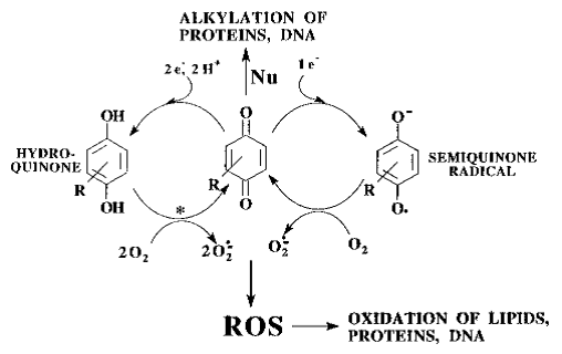
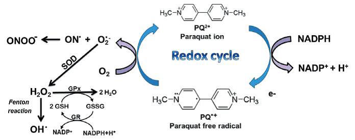
Figure: paraquat RedOx cycle (Blanco-Ayala et al., 2014).

Rotenoids, such as rotenone, often used as pesticides, are RedOx cycling compounds. They bind to complex I of the electron transport chain within the inner mitochondrial membrane where one electron is abstracted from the rotenoid. The radical thus formed can react with oxygen to recover its initial oxidised form while oxygen is transformed into superoxide anion. Then the rotenoid can once again initiate the cycle and generate more superoxide anions leading to important oxidative stress. Moreover, the complex I is inhibited preventing ATP production (Sherer et al., 2007).

Finally, thiols and disulfides can also be the cause of RedOx cycling using the glutathione system as catalyst. Thiolates (basic form of thiols) are oxidised into thiyl radicals which can initiate RedOx cycling with the corresponding disulphide (Munday, 1989). This mechanism depends on stereo-electronic properties of the thiol. Indeed, tertiary thiols do not react the way described hereabove due to steric hindrance. Furthermore, the reactivity of a thiophenol is lower because the electronic conjugation between the sulfur and the aromatic cycle stabilises the thiolate. If the aromatic cycle has groups with electron-withdrawing effect (e.g. halogens or a nitro group), thiolate will be further stabilised and less reactive. On the contrary, if the cycle has electron-releasing groups (alkoxy for instance), the radical is stabilised and oxidation is fostered. For these particular cases, gluthathione, gluthathione-S-transferase and gluthathione peroxidase are not detoxifying, but they favor these toxicity-generating reactions by catalysing the RedOx cycle (Munday, 1989).

close

4.5 Indirect pro-disruptors

This subcategory is related to substances which are metabolised into molecules disturbing enzymes in an indirect way. The final MechoA is explained in MechoA 5.

A priori this MechoA could be found for many substances. To date, only ethanol has been identified using this MechoA even if the narcosis remains the main MechoA for ethanol. The metabolic oxidation of ethanol into acetic acid consumes NAD+ to produce NADH. Thus, high and chronic exposure to ethanol leads to an impaired equilibrium of the cofactor NADH/NAD+. This, added to the induction of high levels of hepatic CYP2E1 and to the reactivity of the intermediate acetaldehyde, leads to liver cancer development among other deleterious effects (King, 2015a).

close

4.6 Docking pro-disruptors

This subcategory is related to substances which are metabolised into molecules that disturb enzymes, receptors or ion channels by directly docking to their active site. This binding can modulate activity of the protein (inhibition or activation). The MechoA of the metabolic product produced further to the reaction is explained in MechoA 6.

Small organo-halogenated compounds, some nitriles and diphenylamines are the main examples of MechoA 4.6.

Trichloroethylene, in the same way as other small organo-halogenated compounds, is metabolised into highly toxic compounds in mammals. The major metabolic pathway is the oxidation into chloral with cytochrome P450, then the reduction into 2,2,2-trichloroethanol (Brüning and Bolt, 2000) (see figure below).


Figure: metabolism of trichloroethylene (Brüning and Bolt, 2000).

The MechoA of 2,2,2-trichloroethanol is related to ion channel modulators (MechoA 6.6). This compound can bind to GABA receptors controlling chloride channels involved in the action potential generation of neurons. Dichloromethane is oxidised in mammals leading to carbon monoxide production (ATSDR, 2014)). The MechoA of carbon monoxide belongs to MechoA 6.9, competing with oxygen to bind hemes and it leads to oxygen deprivation in cells.

Some nitriles can be hydrolysed at the alpha carbon by CYP450 to generate a cyanohydrine. This reactive compound spontaneously releases a cyanide anion on one hand, and an aldehyde or a ketone on the other (Grogan et al., 1992). The cyanide anion (MechoA 6.9) inhibits cytochrome c oxidase, which is the 4th complex within the mitochondrial electron transport chain, thus uncoupling this process from ATP production and generating oxidative stress (Way et al., 2007).

Finally, diphenylamine is another particular case because it can be partially metabolised into indophenol in some mammals (IPCS Inchem, 1998). Indophenol may also inhibit the mitochondrial electron transport chain while the molecular structure of the parent compound does not provide any indication of excess toxicity (Enoch et al., 2008). So far, we have not identified other substances as having similar properties to those defined within this MechoA sub-class.

close

5 Indirect enzyme disruptors

This general MechoA class is dedicated to molecules which indirectly disrupt the operation of enzymes, receptors or ion channels. In this case, xenobiotics do not directly bind to affected proteins, but rather modify their environment. Enzyme function can be disrupted by pH, electrical potential, oxidative power of cofactors, etc.

close

5.1 Oxidative phosphorylation uncouplers

This mechanism is related to the disruption of a pH gradient across the inner mitochondrial membrane. This gradient is generated by the electron transport chain consisting of several membrane enzymes ultimately reducing molecular oxygen into water. The proton gradient is essential for the functioning of ATP-synthase which catalyses ATP production from ADP and inorganic phosphate while transporting one proton to internal matrix.

Some substances are able to transport protons to the internal matrix because they can cross the inner mitochondrial membrane in protonated form (acid form) in one direction and in deprotonated form (basic form) in the other direction. These substances thereby decrease the proton gradient that the electron transport chain is generating, until ATP-synthase can no longer function, and no more ATP is produced. This is called oxidative phosphorylation uncoupling. (Escher et al., 1999).

Organic substances concerned by this mechanism are acidic phenols and all our examples are currently limited to these.

A phenol is considered acidic when there are electron-withdrawing groups pulling the electrons away from the oxygen atom. Consequently, the oxygen will more easily release its proton thus forming a phenolate with negative charge stabilised by electron-withdrawing groups. Under normal conditions, the pH of the inner mitochondrial matrix is around 8 while it is 6.6 in the intermembrane space (Berg et al., 2002; Casey et al., 2010). Therefore, we assume the optimal pKa for a phenol to be an oxidative phosphorylation uncoupler is between 6.6 and 8. It should be noted that certain authors affirm the pKa should be less than 6.5 (Schultz, 1987) to achieve uncoupling.

In order to achieve significant uncoupling, the xenobiotic has to meet several conditions:

- sufficient hydrophobicity,

- pKa within specific limits,

- a certain level of steric hindrance.

All these factors make the uncoupling effect of phenols, and thus their toxicity, difficult to quantify.

Some substances are known to directly interact with the active site of one complex in the electron transport chain thereby disrupting oxidative phosphorylation. In this case, due to direct interaction of the xenobiotic and the complex, the uncoupling effect fall under MechoA 6.

close

5.2 Acids and bases

This mechanism is dedicated to acids with pKa < 5 and bases with pKa of conjugated acid > 9.

Most non-polar chemical species can pass the cell membrane by passive diffusion, but ionic compounds cannot. For acids and bases, at an extracellular pH of around 7.4 and a cytosol pH of 7.2 (Casey et al., 2010), most of these molecules are ionised. Only a small portion of acids and bases exists in neutral form in both compartments. After a neutral molecule has crossed the membrane, it will release or catch electrons in the cytosol to return to its ionised form, driven by the acid-base equilibrium (see figure below).


Figure: example of cell acidification by acids.

In the figure, protonated acids (red) are neutral and can cross the membrane. When arriving in the cytosol, each molecule releases a proton which is recovered by a water molecule (blue) thereby increasing the acidity of the cytosol. Note that, after an acid molecule has passed into the cell, another acid in the extracellular medium becomes protonated because of the acid-base equilibrium (blue). We can assume acidification can also happen in other compartments leading to general disruption of cell functions. This mechanism is partly limited by proton pumps which regulate cell pH. Nevertheless, proton pumps can be overwhelmed when acid or base concentrations become too high.

An example of this is the antimalarial effect of quinine and analogous that has recently been explained by this basification mechanism (Vallabh Minikel, 2016). The author explained that Plasmodium falciparum, the parasite responsible for the disease, is not able to degrade hemes which accumulate in acidic compartments where they spontaneously crystallise. The basification of this compartment by quinine causes heme to be released. Furthermore, free hemes are highly toxic for the parasite because they catalyse oxidation reactions in an uncontrolled and unspecific way leading to radical production. The same author explains that bases, like quinine, can diffuse in the neutral form into more acidic compartments and capture protons. We can assume the MechoA of bases is due to basification of critical cell compartments like lysosomes. In this case proteases would be inhibited. We therefore attribute the cutaneous corrosivity of bases (e.g. pure bases or solutions with pH > 11) to this basification effect.

In parallel, the MechoA of acids must be similar to that of bases except that the critical targets would be the basic compartments such as the inner mitochondrial matrix.

close

5.3 Others

Others MechoAs may exist which involve an indirect disruption of enzyme functions. The only one that has been clearly identified up to now is the MechoA related to ethanol. Ethanol is oxidised to acetaldehyde, then to acetic acid during its metabolism. The enzymes dealing with these transformations reduce NAD+ into NADH as in the Krebs’ cycle. Therefore, in the case of high exposure to ethanol, glycolysis rate and fatty acid oxidation may be decreased by a lack of NAD+. This would lead to an excess of fatty acids in blood and in liver potentially causing hepatic steatosis or even liver cirrhosis (King, 2015a). In such cases indirect disruption of the whole enzymatic system may occur due to a lack or an excess of the common cofactor (NAD+/NADH).

close

6 Direct docking disruptors

This general MechoA class is includes the largest number of MechoA subcategories and is related to any direct interaction with an enzyme, a receptor or an ion channel (called active protein here).

The most well-known MechoAs within direct docking disruption are: acetylcholine esterase inhibition, nicotinic or muscarinic acetylcholine receptor binding, ion channel binding (calcium, sodium, chloride, etc.), opioid receptor binding, heme binding and hormonal receptor binding (oestrogen, androgen, thyroid).

The binding of a xenobiotic to an active protein can be localised at the active site of the enzyme, to an allosteric site of an enzyme, an ion channel or a receptor, or even to the main binding site of a receptor.

close

6.1 Acetylcholine-esterase inhibitors

This mechanism is responsible for the disruption of the acetylcholine (ACh)-dependant nervous transmission. ACh is a neurotransmitter which triggers the opening of ions channels, which are either the receptors themselves for nicotinic receptors or separate ions channels for muscarinic receptors. The ion flux leads to depolarisation of the postsynaptic cell thus initiating the electric signal through it. Once the transmission of the signal is achieved, the activity due to ACh must be stopped in order to close ions channels and the cell to recover its resting state. The acetylcholinesterase (AChE) hydrolyses the ACh into choline and acetate in the synaptic cleft. AChE is therefore critical to ensure that the nervous transmission is executed. The choline molecules produced with AChE activity are recycled by transporting them back to the neurone which initially secreted the ACh.

The inhibition of AChE rapidly leads to lethal effects for animals by arresting respiratory muscle functions (Elersek and Filipic, 2011; Fukuto, 1990). This mechanism is used by nerve agents for chemical weapons (e.g. VX, sarin, tabun, etc.). This is also a common mechanism of action used by various insecticides like azinphos-methyl, methomyl, aldicarb, malathion, etc.

At a molecular level, the hydrolysis of ACh includes several steps: binding of ACh to the serine residue at the AChE active site, elimination of choline and elimination of acetate.

Inhibition of AChE is possible by imitating ACh binding to serine as presented in the figure below. As expected, the leaving group is eliminated in the second step. However, the group attached to serine is barely eliminated compared to ACh (Elersek and Filipic, 2011).

Organophosphorus compounds with structures shown in the figure are known to have strong capacities to inhibit AChE. There may be other less well known enzymatic targets of organophosphorus compounds as suggested by (Elersek and Filipic, 2011). Carbamates are another chemical family with good candidates for AChE inhibition (Fukuto, 1990).

Some structure-activity relationships for these compounds have been established. Ester reactivity (for organophosphorus) and the strength of the leaving group (for carbamates) have been identified as having a strong impact on the inhibition efficiency. Nevertheless, reactivity is not the only parameter leading to inhibition. Steric hindrance also plays an important role. For example, carbamates with a good leaving group such as a phenoxy or an oxime must have an adequate three-dimensional structure corresponding to the form and polarity of AChE active site (Fukuto, 1990).

This MechoA occurs only for organisms with neurons, i.e. most animals with a few exceptions. However, substances with MechoA 6.1 (notably organophosphorus compounds) are not necessarily inert for non-animal organisms because they can inhibit other esterases.

close

6.2 Acetylcholine-receptor binders

This mechanism is responsible for the disruption of the nervous transmission by binding the acetylcholine receptors (AChR). A molecule acting through MechoA 6.2 can be an agonist or an antagonist.  Agonists produce the same effect as natural ligands, i.e. AChR activation. Antagonists block the AChR receptor rather than activating it like an agonist.

Note that ACh agonists to AChR produce a similar effect as AChE inhibitors as they activate AChR but are not degraded by AChE, therefore the effect is not stopped, and the post-synaptic cell cannot recover its resting potential.

The best known ACh agonists are nicotine for nicotinic receptors (nAChR) and muscarine for muscarinic receptors (mAChR).

Antagonists are toxic when they are administered alone by blocking nervous transmission. However, they can be used as antidotes to prevent adverse effects occurring due to exposure to AChE inhibitors and AChR agonists.

Atropine and derivatives are typical examples of mAChR antagonists (IPCS Inchem, 2002). Besides coniine is known to block nAChRs (NCBI, 2017; Vetter, 2004). Compounds with a similar structure to coniine can also cause the same effect despite their very simple structure. The efficiency of these antagonists depends on their affinity to the binding site (Committee on Acute Exposure Guideline Levels et al., 2012).

Agonists and antagonists to ACh share common features with ACh (see Figure below). All of them have a bulky moiety with a positive charge (in blue) at a few Angströms distance from an electron-rich H-bond acceptor (in red).


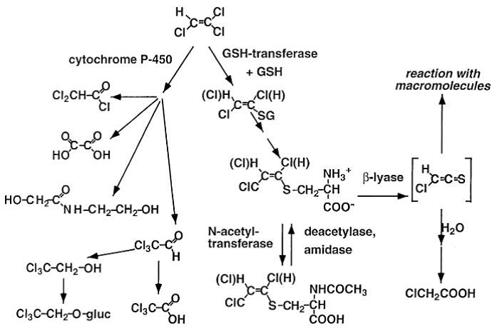
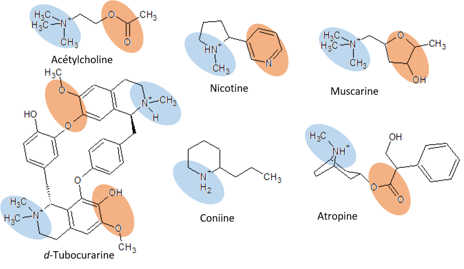
Figure: Molecular structures of acetylcholine and some agonists and antagonists.

Tubocucarine is also an antagonist of ACh for nAChR and it contains 2 equivalents of ACh (King, 2015b). Indeed, there are 2 binding sites for ACh in AChR which have to be occupied to open ion channel of the receptor (Droual, 2011). The reason why a molecule will have an agonist effect or antagonist effect is not yet totally understood.

Nota Bene. This mechanism only concerns organisms with neurons which use ACh as their neurotransmitter (with AChRs). Basically, these are animals with some exceptions.

close

6.3 Dopamine transport disruptors

Dopamine is a neurotransmitter which is involved in other neuronal connections than acetylcholine. As for acetylcholine, the functioning of the nerve transmission needs dopamine to be released at the synaptic cleft in order to activate a G Protein-Coupled Receptor. The neurotransmitter is then recycled once it returns to the presynaptic neurone (Calipari and Ferris, 2013; King, 2015b).

Amphetamine and similar compounds modify these mechanisms.

Amphetamines force dopamine release in the synaptic cleft. At the same time, it inhibits the transport of dopamine back to the cell leading to an “overdose” of dopamine in the intercellular space and thereby, continuous excitation of the postsynaptic neurone (Calipari and Ferris, 2013).

This mechanism of action concerns only organisms with dopamine-dependant neurones, basically animals with some exceptions (Barron et al., 2010). Note that dopamine has also been identified in plants, but in this case the substance has a different role (Kulma and Szopa, 2007).

close

6.4 Metal chelators

For most organisms, metals are used to a great extent for enzyme function, inside their active sites, to catalyse chemical reactions. For instance, an iron ion is present in the centre of cytochrome P450 heme, and metalloproteases use a magnesium or zinc ion to catalyse hydrolysis reaction. On the other hand, metal ions can play a critical role in cell signalling (especially calcium ions) or they are used to create the electric action potential (sodium and potassium). Metal chelation is the formation of a stable complex between a molecule and a metal ion. Some xenobiotics have the ability to chelate metals thus competing with enzymes or disrupting cell signalling. These compounds can either share the metal chelation inside the active site of the enzyme, either extract the metal ion from the enzyme to form an independent complex. In both cases, the enzyme is inactivated.

In order to have the capacity to chelate metals, the molecule must have 2 or more negative partial charges at the right distance corresponding to the diameter of the target metal. For instance, beta-dicarbonyls are efficient iron chelators and degrade cytochrome P450, provided the two carbonyls can get into a parallel position (O’Donoghue, 2001). Calcium can be chelated by oxalate forming crystals which accumulate and block the blood circulation (Wiley-VCH Verlag, 2002b).

Some organisms have protecting proteins called metallothioneins, which can chelate metal ions to control the free concentration and these are useful as a protective mechanism in the case of exposure to heavy metals, for example. However, gene expression of metallothioneins is finely regulated. Therefore, high exposure to xenobiotic metal chelators is deleterious for the cell (Petering and Fowler, 1986).

close

6.5 Photosystem II electron transport inhibitors

This mechanism is similar to RedOx cycling (MechoA 4.4) with the main difference that instead of blocking mitochondrial electron transport chain, substances with MechoA 6.5 block the electron transport chain of plant photosystems.

Several urea derivatives inhibit D1 protein in photosystem II (van Rensen, 1982) preventing electron transmission from water oxidation to other proteins of the chain which reduce NADP+ to NADPH. Besides this, the electron transport chain creates a proton gradient between the stroma and the thylakoid lumen which is used by ATP synthase to produce ATP. By blocking electrons at the level of the D1 protein, no more NADPH or ATP is produced.

From https://www.khanacademy.org/science/biology/photosynthesis-in-plants/the-light-dependent-reactions-of-photosynthesis/a/light-dependent-reactions 

Bromacil, Diuron, Atrazine and other triazines are typical examples of compounds which inhibit D1 protein in photosystem II.

close

6.6 Ion channel modulators

This MechoA is related to substances which modify activity of ion channels or ionotropic receptors except for nicotinic acetylcholine receptors (nAChRs) for which a dedicated MechoA class (MechoA 6.2) exists.

Substances with MechoA 6.6 may disrupt the functioning of receptors to γ-aminobutyric acid (GABA), a neurotransmitter. There are two types of GABA receptors, GABAAR and GABABR, which are directly (GABAAR) or indirectly (GABABR) responsible for the influx of chloride ions. Activation of GABAergic neurons by anticonvulsant, anxiolytic, sedative and hypnotic drugs leads to antidepressant effects (Olsen and DeLorey, 1999).

2,2,2-trichloroethanol has a GABAergic activity by binding to GABA receptors which then become more sensitive to GABA.

Neurons also have G protein-coupled opioid receptors which trigger the opening of channels for potassium efflux and calcium influx. The activation of opioid receptors leads to the reduction of action potential and nerve signal propagation.

Morphine and derivatives are the best known opioids with MechoA 6.6 (Chahl, 1996).

Some substances prevent the closing of sodium channels in neurons thus prolonging their excitation.

These are basically DDT (dichlorodiphenyltrichloroethane) and analogues like methoxychlor and pyrethroids like resmethrin, cypermethrin or tefluthrin, etc. (figure below).


Figure: Structure of DDT derivatives and pyrethroids derivatives.

Besides having impact on sodium channels, type II pyrethroids also inhibit the opening of GABAergic chloride channels and Ca-Mg-ATPase thus resulting in a modification of calcium concentration in the neuron (Coats, 1990). Moreover, various substances can modulate GABAergic calcium channels which have several different binding sites with a broad range of potential modulations (see figure below).

Figure: GABA receptor and binding sites (Olsen and DeLorey, 1999).

The binding site of 2,2,2-trichloroethanol and ethanol is uncertain (Olsen and DeLorey, 1999). It is postulated that pyrethroids bind to the picrotoxin site (Coats, 1990). Chlorinated alicyclic insecticides also bind to the picrotoxin site thus preventing the channel opening and the neuron recovering its resting state. These insecticides would therefore cause death by respiratory arrest (Coats, 1990). They have various structures with a non-aromatic cycle (i.e. alicyclic) and several chlorine substituents as a common point. Some examples are presented in the figure below.

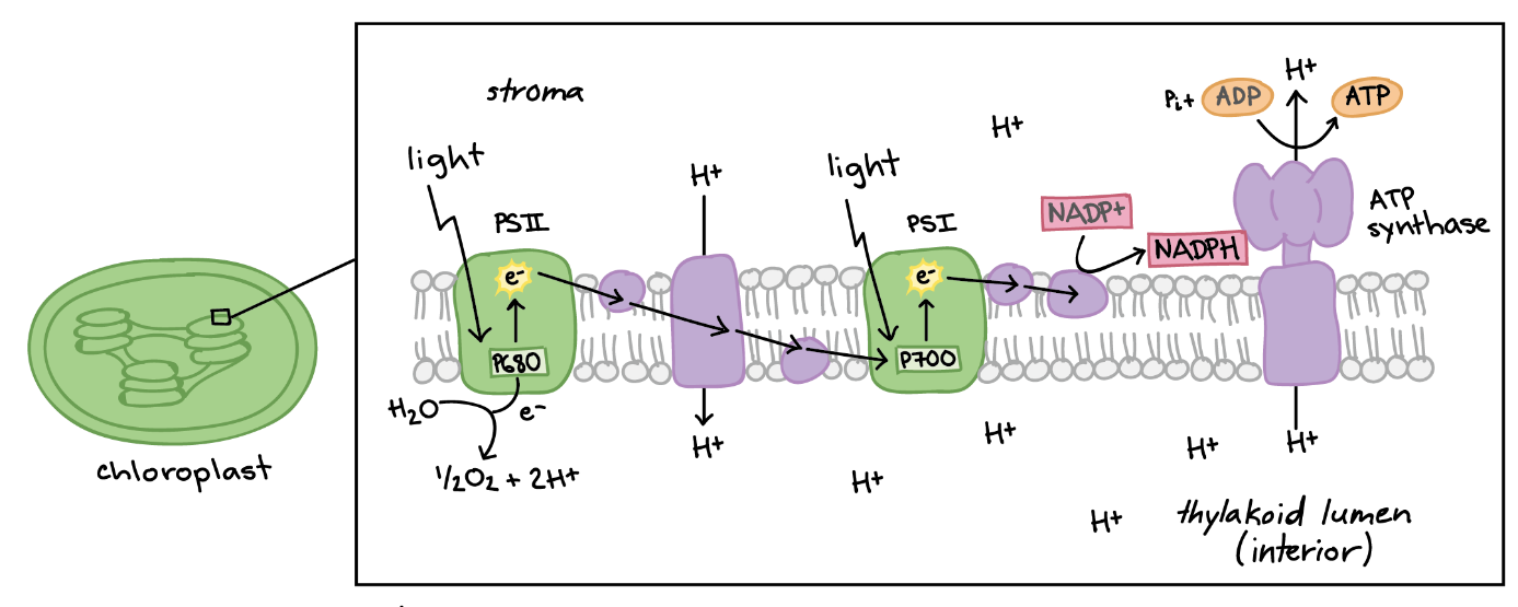
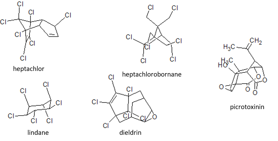
Figure: chlorinated alicyclic insecticides and picrotoxinin.

Caffeine is another example of substance which has MechoA 6.6 because it binds to the GABA receptor at benzodiazepine sites. However, this mechanism is minor for caffeine (and methylxanthines, in general) which modulates several calcium channels by activating or disactivating them depending on the dose. Furthermore, caffeine is an antagonist of adenosine receptors leading to repression of the spontaneous neuronal electric activity, synaptic transmission inhibition and neurotransmitter release (Nehlig et al., 1992). Caffeine is also believed to disrupt calcium regulation in plants, but these mechanisms are not well studied (IAC Publishing Labs).

close

6.7 Mitochondrial electron transport chain inhibitors

This MechoA is related to substances which inhibit proteins involved in the electron transport system of mitochondria present in every animal cell. When one of these proteins is inhibited, the whole chain is disrupted, and the proton gradient is no longer maintained preventing ATP production.

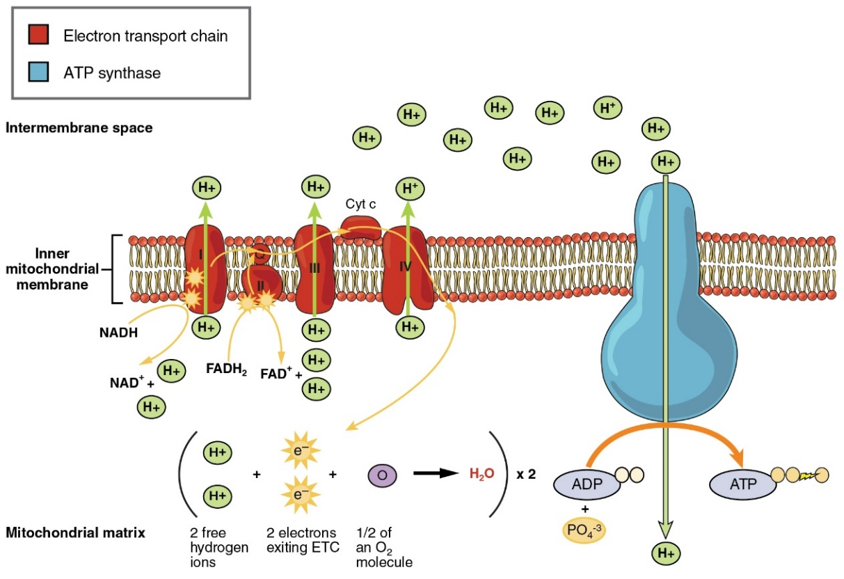
Figure: Coupling of ATP synthase and the electron transport chain with the proton gradient. Taken from Anatomy & Physiology (OpenStax, 2014).

The best-known examples are rotenone (and rotenoids in general) which inhibit complex I, cyanide ion which inhibits complex IV.

close

6.8 Endocrine disruptors

This MechoA concerns substances that disturb the production, transport or metabolism of hormones, or that interact with their receptors, either as agonist, antagonist, or any other mechanism that alter the number of hormone receptors or their activity by docking. Currently there is focus particularly on the Estrogen, Androgen, Thyroid and Steroidogenesis (EATS) disruption although many other enzymes and receptors could ultimately be implicated in endocrine disrupting pathways.


Examples of this MechoA are ethinylestradiol (EE2), tamoxifen (Estrogen disruption), DDT ,flutamide (Androgen disruption), benzophenones, amitrole (Thyroid disruption), phthalates and epoxiconazole (Steroidogenesis disruption).


The prediction of this endocrine disruptor MechoA is provided by KREATiS using a battery of tests as part of its specific expert services. Please send a mail to contact@kreatis.eu for further details.

close

6.9 Others

There are various other MechoAs involving specific interactions with an active protein which are sometimes specific to one particular molecule (e.g. amanitin). These mechanisms are listed below without detailing them. There are of course other MechoAs which are not in this list, notably MechoAs of active pharmaceutical ingredients. At KREATiS, we are working daily to improve our understanding of specific MechoAs and to include them in the MechoA Classification Scheme.

Non-exhaustive list of other specific MechoAs:

  1. Inhibition of cyclooxygenases (COX-1 and COX-2) (e.g. salicylic acid and derivatives)
  2. MechoAs of specific herbicides (e.g. glyphosate, trifluralin)
  3. Glycine antagonism (e.g. strychnine)
  4. Aryl hydrocarbon receptor binding (e.g. dioxins, PCBs and polychlorodibenzofurans)
  5. Inhibition of oxygen transport by haemoglobins (e.g. carbon or sulfur monoxide, cyanide ion)
  6. Inhibition of DNA transcription by binding RNA polymerase (e.g. amanitine)